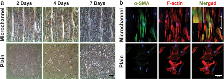
FIG. 4.
(a) Microscopic images of vascular smooth muscle cells grown on microchanneled and plain gelatin substrates: controlled topography induces elongated and aligned SMCs on microchannels and plain substrate allows random elongation of actin fibrils. (Scale bar = 200 μm). (b) Alpha smooth muscle actin (α-SMA, green), F-actin (red), DAPI (nuclei, blue), and merged actin expression in SMCs cultured in the microchanneled and plain substrate after 7 days (white arrow indicates fiber orientation; scale bar = 100 μm). Images by Tijore et al.115 with permission from Springer. Color images are available online.

