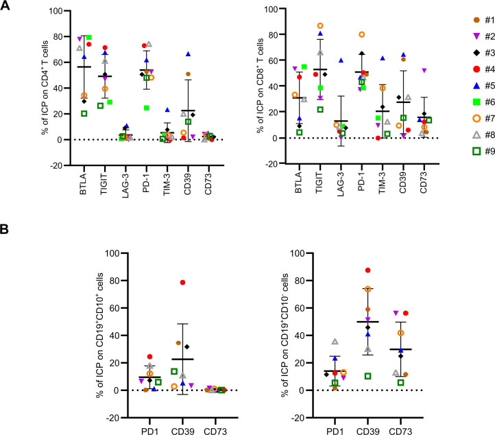
Figure 5.
FL-PDLS immune checkpoint characterization. 10 FL-PDLS from nine different FL patients at day 3 of culture were pooled and the percentage of ICP was analyzed by flow cytometry. (A) Percentage of BTLA, TIGIT, LAG-3, PD-1, TIM-3, CD39, CD73 on CD4+ and CD8+ T cells. (B) Percentage of PD-1, CD39, CD73 on tumorous cells (CD10+CD19+) and healthy B cells (CD10−CD19+). FL-PDLS, follicular lymphoma-patient-derived lymphoma spheroid; ICP, immune checkpoint.

