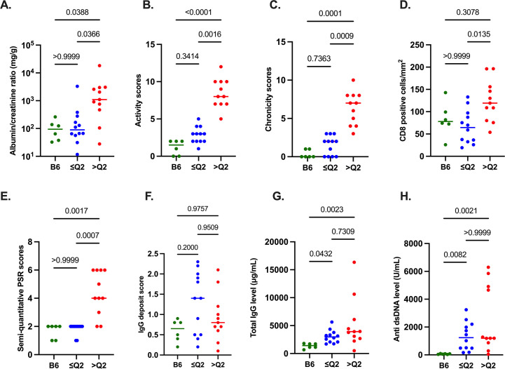
Figure 5.
Association of p16Ink4a staining with renal disease in a second (ageing) cohort: 8–12 month-old mice (6 B6, 23 B6.Sle1.2.3 mice). (A–C) Renal disease parameters: urine albumin/creatinine ratio (mg/g), histopathological activity and chronicity scores on H&E-stained FFPE kidney sections (scale: 0–12). (D,E) Quantification of anti-CD8 staining (positive cells/mm2) and semi-quantitative PSR staining scores (scale: 0–6) on serial FFPE kidney sections (with p16Ink4a). (F) Semiquantitative evaluation of glomerular IgG deposits in frozen OCT kidney sections. (G,H) Systemic disease parameters: total IgG (μg/mL) and anti-dsDNA antibody (U/mL) in plasma. Horizontal bars: medians. p values: Kruskal-Wallis and Dunn’s post-hoc tests. ≤Q2: p16Ink4a-low (≤50th percentile), >Q2: p16Ink4a-high (>50th percentile) B6.Sle1.2.3 kidneys. anti-dsDNA, anti-double-stranded DNA; B6.Sle1.2.3, B6.NZMSle1/Sle2/Sle3; FFPE, formalin-fixed paraffin-embedded; OCT, optimal cutting temperature; PSR, Picrosirius red.

