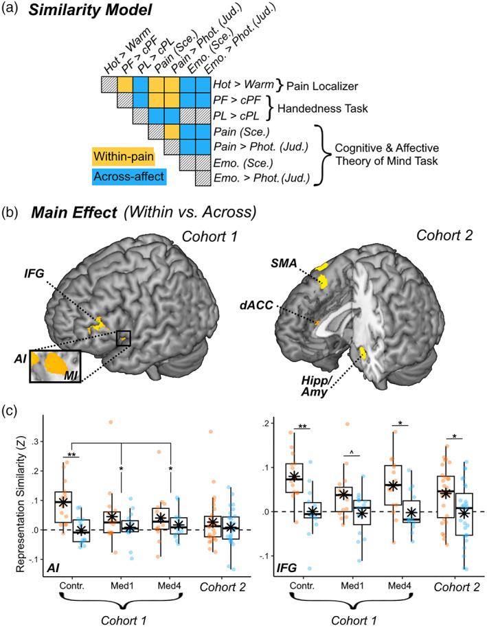
FIGURE 5.

Representational similarity of pain. (a) Schematic matrix of a representation model testing for pain‐specific activity patterns across different tasks in our study. Within the matrix, row and column labels refer to conditions of interest: Hot temperatures from the “Pain Localizer,” Painful (PF) and Negative Painless (PL) images from the “Handedness” task, and Beliefs, Emotions, and Pain Scenarios & Judgments from the “Cognitive and Affective Theory of Mind” task. Each matrix cell indicates the putative representation similarity between two conditions: yellow cells concern pairs of pain conditions (within‐pain), blue cells concern pairs of a painful and emotional painless conditions (Across‐affect), and black‐striped cells refer to pairings of no interest. (b) Surface brain rendering of regions displaying significant main effects in their sensitivity to pain‐specific information (within‐pain vs. across domain). Detailed coordinates are listed in Table S12. (c) Activity parameters extracted from the highlighted region are plotted across groups and cohorts, with boxplots and individual data. Amy, amygdala; Contr., controls; dACC, dorsal anterior cingulate cortex; Hipp, hippocampus; IFG, inferior frontal gyrus; Med1 & Med4, university students enrolled at the first/fourth year of medicine; MI & AI, middle & anterior insula; SMA, supplementary motor area. “***,” “**,” and “*” refer to significant effects differences from non‐parametric rank tests at p < .001, p < .01, and p < .05 respectively.
