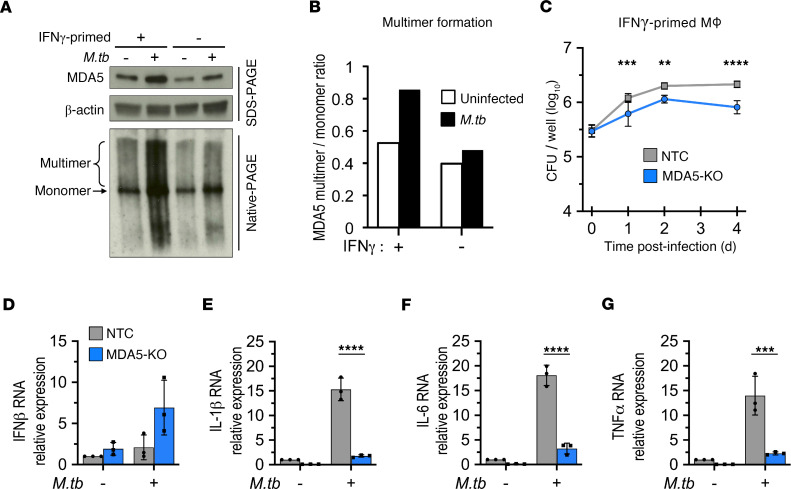
Figure 6. MDA5 is activated during M. tuberculosis infection and promotes intracellular bacillary growth in primed human macrophages.
(A) IFN-γ–primed or resting human monocyte-derived macrophages were infected with M. tuberculosis (MOI of 1:10) for 24 hours. Whole-cell lysates were subjected to native or SDS-PAGE followed by immunoblot analysis. Data are representative of 1 independent experiment. (B) Densitometric analysis was used to quantify band intensities in A presented as a ratio of MDA5 multimer to monomer. (C) Growth kinetics of M. tuberculosis (MOI of 1:5) in PMA-differentiated and IFN-γ–primed THP-1 cells (NTC and MDA5-KO). Data are the mean CFU ± SD for each time point (n = 6 biological replicates from 2 independent experiments). (D–G) PMA-differentiated and IFN-γ–primed THP-1 cells (NTC and MDA5-KO) were infected with M. tuberculosis (MOI of 1:5) for 24 hours. RNA levels of IFN-β (D), IL-1β (E), IL-6 (F), and TNF-α (G) were measured by reverse transcriptase qPCR and presented as fold-change relative to NTC no-infection control. RNA levels were normalized to PolR2A expression (mean ± SD, n = 3 biological replicates from 3 independent experiments). **P < 0.01, ***P < 0.001, ****P < 0.0001 by 2-way ANOVA with Tukey’s posttest.

