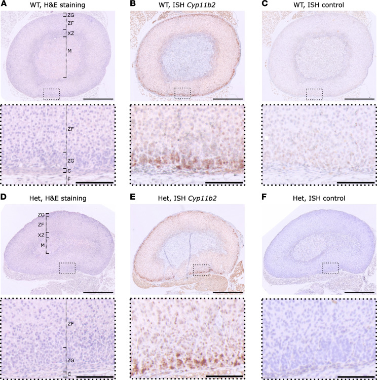
Figure 3. Adrenal histology and Cyp11b2 ISH of Cacna1dIle772Met/+ and WT.
(A and D) H&E staining of FFPE adrenal sections from 14-week-old female WT (A) or Cacna1dIle772Met/+ mice (Het, D). F, fat; C, capsule; ZG, zona glomerulosa; ZF, zona fasciculata; XZ, x-zone, M, medulla. (B and E) ISH of WT or Het mice. A probe for the detection of Cyp11b2 mRNA was used. Staining is confined to the ZG. (C and F) Controls for ISH without any probes are shown. The stainings shown in this figure are representative for 8 WT (6 female, 2 male) and 6 Het (3 female, 3 male). Scale bars: 500 μm (overview, top) or 100 μm (magnification, bottom).

