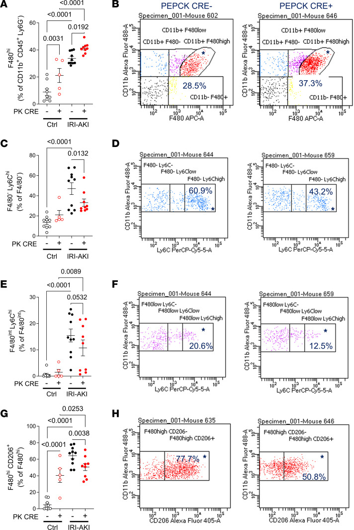
Figure 10. Flow cytometric analysis of renal monocyte/macrophages in PTEC DN RAR mice.
Kidneys were harvested from PTEC DN RAR mice 3 days after bilateral IRI-AKI. (A and B) F4/80hi cells (kidney-resident macrophages). (A) F4/80hi cells as the percentage of gated CD11B+CD45+Ly6G– cells. (B) CD11B and F4/80 expression charts in PEPCK Cre+ and Cre– mice after IRI-AKI (percentage in gated area indicated). (C–F) Ly6Chi cells (inflammatory monocyte/macrophages). (C and E) F4/80– (infiltrating monocytes) Ly6Chi cells and F4/80int (BM-derived macrophages) Ly6Chi cells, as the percentage of gated F4/80– and F4/80int cells, respectively. (D and F) CD11B and Ly6C expression charts. (G and H) CD206/mannose receptor expression (an M2 activated macrophage marker). (G) CD206+ cells as the percentage of gated F4/80hi cells. (H) CD11B and CD206 expression charts. A, C, E, and G used 1-way ANOVA, with P < 0.05 considered significant; q values shown for between-group comparisons corrected for repeated testing.

