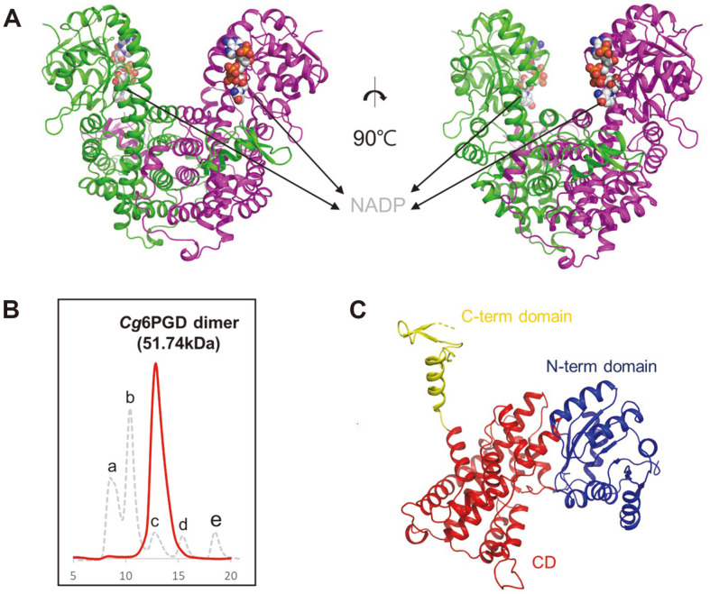
Fig. 2. Overall structure of Cg6PGD.
(A) Dimeric structure of Cg6PGD. The dimeric structure is shown as a cartoon model, and the two chains are distinguished with different colors : green and magenta, respectively. NADP is presented as a gray-colored sphere model. (B) Size-exclusion chromatography of Cg6PGD. a is a void peak, b is ferritin (440 kDa), c is aldolase (158 kDa), d is ovalbumin (44 kDa) and e is ribonuclease (13.7 kDa). Cg6PGD is eluted as a dimer. (C) Domain classification of Cg6PGD using a cartoon model of the monomer. The N-term domain is indicated in blue, the CD is indicated in red, and the Cterm domain is indicated in yellow.

