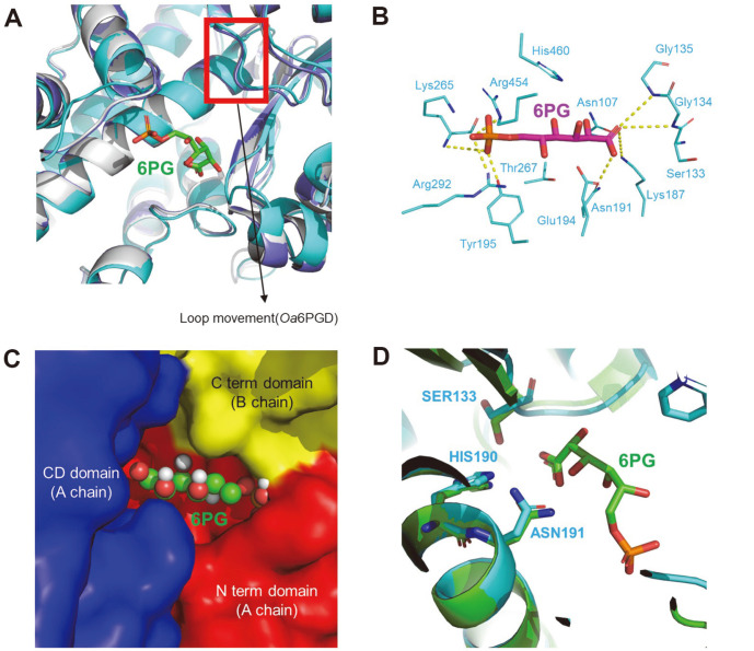
Fig. 3. Substrate(6PG)-binding model of Cg6PGD.
(A) Structure comparison between Oa6PGD_apo, Oa6PGD_6PG and Cg6PGD_apo. All structures are presented as cartoon diagrams. The structures are depicted in gray (Oa6PGD_apo), purple (Oa6PGD_6PG) and cyan (Cg6PGD). (B) Docking simulation of Cg6PGD with 6PG. Hydrogen bonds between the residues and 6PG are expressed, with the exception of water-meditated hydrogen bonds. 6PG is presented as a magenta-colored stick model, and the substrate-binding residue is presented as a cyan-colored stick model. All residues are labeled. (C) Domains associated with the binding of 6PG. (D) Catalytic residues of Cg6PGD and Oa6PGD. Cg6PGD is presented as a cyan-colored cartoon model, and Oa6PGD is presented as a green-colored cartoon model. The catalytic residues are presented using sticks and labeled.

