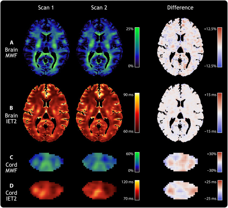
Fig. 2. Reproducibility of MWI metrics in brain and spinal cord.
Individual slices of the (A) brain MWF, (B) brain geometric mean of IET2, (C) spinal cord MWF, and (D) spinal cord IET2 for a single healthy participant. Columns show results from two separate CALIPR MWI exams, aligned in the subject’s structural image space (T1-weighted for the brain and T2*-weighted for the cord), along with their voxel-wise difference maps.

