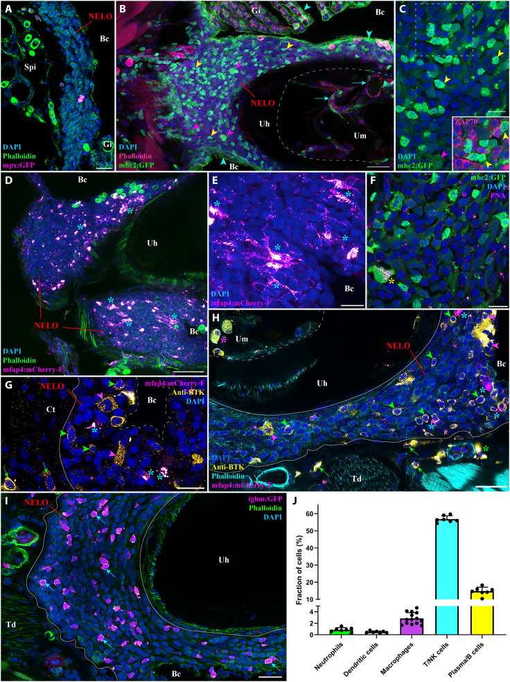
Fig. 3. NELO immune cell populations.
(A) Adult mpx:GFP zebrafish NELO cryosection, in which neutrophils are fluorescent (magenta hot). (B) Adult mhc2:GFP zebrafish cryosection, in which mhc2-expressing cells are fluorescent (green). The expression of MHC2 is observed in the epithelial cells (cyan arrowheads), large cells inside NELO (magenta arrowheads), and within the marrow of the urohyal bone (cyan arrows). (C) Zoom inside NELO of a mhc2:GFP fish highlighting large (magenta arrowheads) and small (yellow arrowheads) positive cells. The latter being negative for anti-ZAP70 labeling (cherry). (D and E) Adult mfap4:mCherry-F zebrafish NELO cryosection in which macrophages are fluorescent (magenta hot, cyan stars). (F) Cryosection of an adult mhc2:GFP zebrafish NELO stained with peanut agglutinin lectin (magenta hot) to reveal dendritic cell (yellow star). (G and H) Anti-Bruton’s tyrosine kinase (BTK) labeling (yellow hot) of mfap4:mCherry-F adult zebrafish cryosections revealed macrophages (magenta hot, cyan stars), BTK-positive B cells (green arrowheads) and BTK-positive plasma cells (magenta arrowheads) in NELO. BTK-positive cells were also found in the connective tissue surrounding NELO (green arrows), around endothelial structures (green stars), and within the marrow of the urohyal bone (magenta star). (I) The presence of B cells in NELO was confirmed using ighm:GFP zebrafish, in which a subtype of B cells that express immunoglobulin M is fluorescent (magenta hot, cyan arrows). (J) Quantification of the different immune cell populations found in NELO counted from at least seven single-cell layer images originating from at least three fish. Annotation: Um, Urohyal marrow. Scale bars, 50 (D), 30 (B), 20 (A, H, and I), and 10 (C and E to G).

