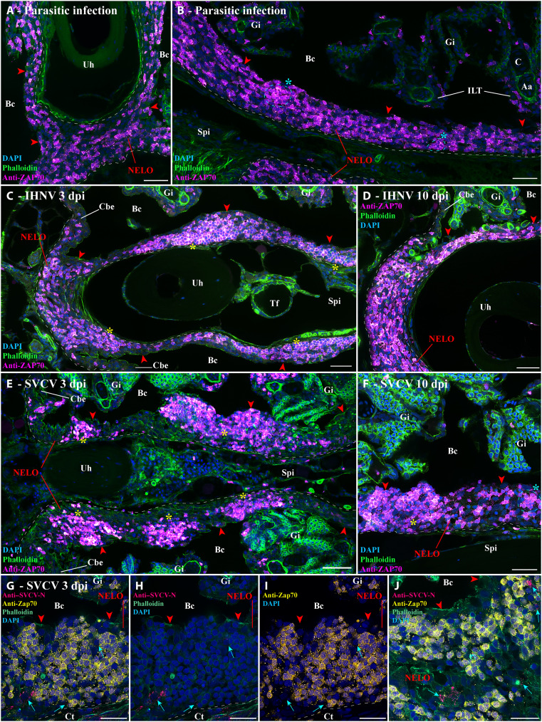
Fig. 5. Structural response of NELO to viral and parasitic infections.
(A and B) Cryosections displaying NELO (red arrowheads) in adult zebrafish naturally coinfected with three parasite diseases (P. neurophilia, P. tomentosa, and M. streisingeri) labeled with anti-ZAP70 antibody (magenta hot). The distribution of ZAP70-positive cells in NELO appears more heterogeneous than in uninfected fish. In addition, some the labeled cells formed small clusters (cyan stars). (C and D) Cryosections from adult zebrafish bath-infected for 24 hours with IHNV. After the apparition of more prominent aggregations of ZAP70-positive cells at 3 dpi (yellow stars) (C), the distribution of T/NK cells reverted to a more homogeneous state by 10 dpi (D). (E and F) Cryosections from adult zebrafish bath-infected for 24 hours with SVCV. NELO displayed notable aggregations of T/NK cells into distinct clusters at 3 dpi (yellow stars) (E). A week later, NELO displayed both large (yellow star) and small clusters (cyan star) of ZAP70-positive cells (F). (G to J) Cryosections from zebrafish 3 day after SVCV infection co-labeled with anti-ZAP70 antibody (yellow) and anti-SVCV-N antibody (cherry), revealing cells loaded with viral material (cyan arrows) neighboring large clusters of ZAP70-positive cells. Annotations: dpi, day postinfection; IHNV, infectious hematopoietic necrosis virus; SVCV, spring viremia of carp virus. Scale bars, 50 (E), 30 (A to D and F), and 20 μm (G to J).

