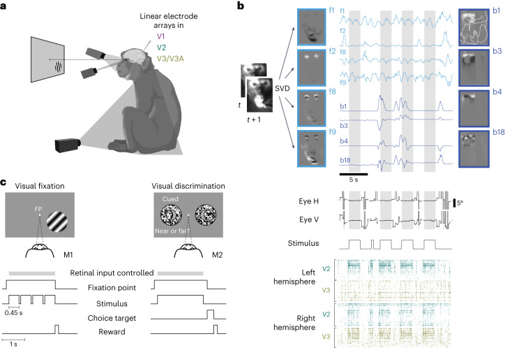
Fig. 1. Monitoring spontaneous body movements during task performance in macaque monkeys.
a, The set-up. The animals performed a visual task while extracellular activity in their visual cortex was recorded and the animals’ body, face and eye movements were monitored via video, with one camera directed at the body, one at the face and a video-based eye tracker. b, Movements recorded by video (example from M2) were decomposed (SVD), generating multiple components of face and body movements that map on to, for example, movements of the mouth (face component 1 (f1)), eye blinks (f2), combinations of face parts (f8 and f9) and combinations of hand, arm, leg and body movement (body components b1, b3, b4 and b18; outline of the monkey body shown in b1; grayscale shows normalized components; traces show normalized temporal profiles of the video projected on to the components); the middle panels show eye positions and stimulus ON/OFF periods. Dark-gray bands in the eye position traces indicate interrupted eye signals resulting from blinks or eccentric eye positions; light-gray shading marks epochs when the animals fixated and the retinal input was controlled. Bottom, sample spike rasters of simultaneously recorded units in the left and right hemispheres of V2 and V3/V3A. In each row spike times from one unit are shown as vertical ticks. c, Animal M1 performing a visual fixation task and animal M2 performing a visual discrimination task combined with block-wise manipulation of spatial attention. The retinal input was controlled during periods (gray bar) when the animals fixated on an FP at the center of the screen.

