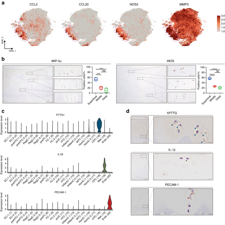
Fig. 2.
Signatures and putative spatial distribution of InflamC, CPC, Mac and EndC. a Expression of selected marker genes (combined for annotation) for InflamC visualized by a feature plot. b Representative immunohistochemistry (IHC) staining for MIP-3α and iNOS in hand articular cartilage tissues (n = 4). Scale bar, left, 100 μm; right, 20 μm. ***P < 0.001, ****P < 0.000 1. c Violin plot of selected marker genes for CPC, Mac and EndC. d Representative IHC staining of hPTTG, IL-1β and PECAM-1. Arrows indicate positive cells in the cartilage tissue. Scale bar, left, 100 μm; right, 10 μm. InflamC inflammatory chondrocytes, CPC cartilage progenitor cells, Mac macrophages, EndC endothelial cells, MIP-3α macrophage inflammatory protein 3 alpha, iNOS inducible nitric oxide synthase, hPTTG human pituitary tumor-transforming gene 1 protein, IL-1β interleukin-1 beta, PECAM-1 platelet endothelial cell adhesion molecule

