
Fig. 1.
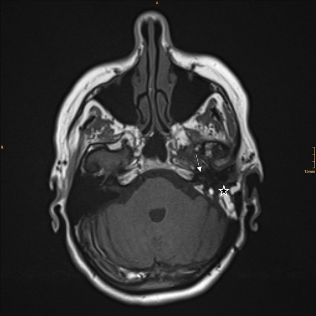
MRI T1, axial, postoperative image. The arrow points at the cochlea, the star is placed in the previous mastoid region showing postoperative changes. This image was rated as Grade 3—excellent image quality
Official websites use .gov
A
.gov website belongs to an official
government organization in the United States.
Secure .gov websites use HTTPS
A lock (
) or https:// means you've safely
connected to the .gov website. Share sensitive
information only on official, secure websites.
MRI T1, axial, postoperative image. The arrow points at the cochlea, the star is placed in the previous mastoid region showing postoperative changes. This image was rated as Grade 3—excellent image quality