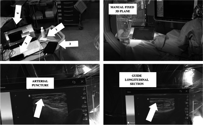
Fig. 4.
Different views of the system. Top left quadrant shows the main hardware components of the transmitter module: (1) ultrasound scanner, (2) video capture card, (3) tablet, and (4) laptop. Top right quadrant shows a plane rendering ultrasound scanner images fixed manually by the doctor over the patient using the button placed on the top right corner of the window (next to the cross button). Bottom-left quadrant shows an arterial puncture assisted by the system. Bottom-right quadrant shows the guide introduced into the artery from a longitudinal section view

