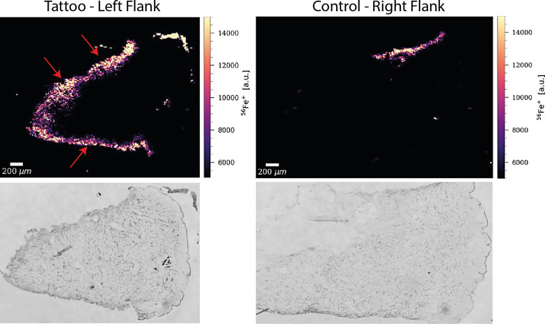
Fig. 2.
Laser ablation-ICP-MS, punch biopsies, and cross-sectional imaging with the visualization of iron (Fe) distribution in the punches (epidermal surface to the right); left column: from the tattoo; right column: reference normal skin from the contralateral side. Red arrows indicate iron contamination of the tissue along the cutting line originating from the punch. Figures below show unstained tissue sections of the biopsy cores.

