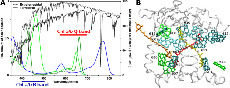
Figure 1.
Spectroscopy and structural arrangement of typical plant photosynthetic pigments. (A) Ultraviolet/visible region absorption spectra of Chls a, b, and BChl a (dark green, light green, and blue, respectively) compared to solar irradiation.20 (B) CP29 antenna complex from Pisum sativum, PDB structure 5XNL.45 Viewpoint along thylakoid membrane plane, looking toward the center of the photosystem II supercomplex. Stromal side at the top and lumenal side at the bottom. Protein gray and transparent, Chls a shown as cyan, Chls b as green (Chl numbering as given in earlier CP29 structures, e.g., 3PL9). Crts are also shown, lutein in yellow, neoxanthin in orange, and violaxanthin in red.

