
Figure 3.
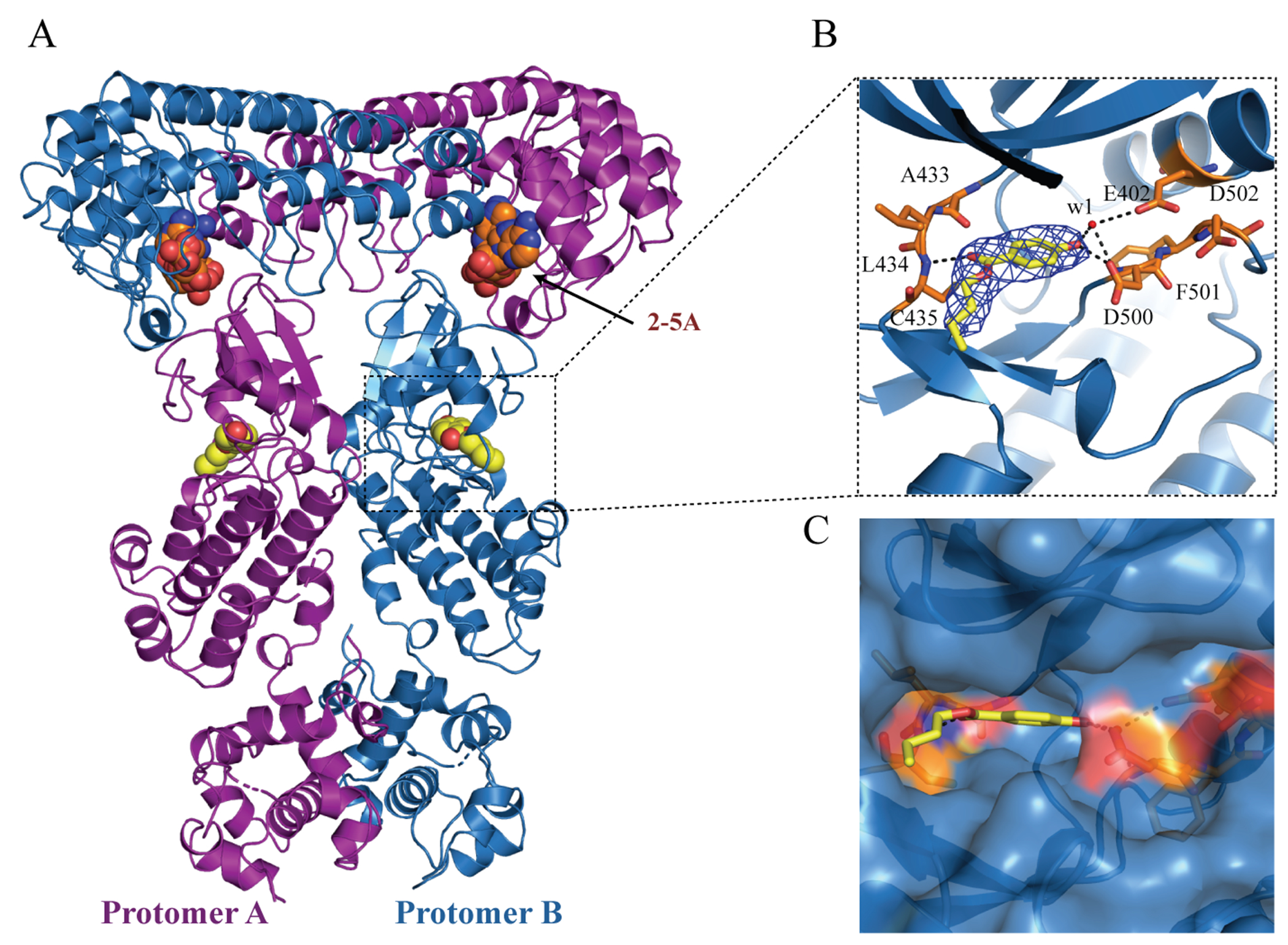
Co-crystal structure of the dimeric P-RNase L in complex with the fragment AC40357. (A) The dimer structure of P-RNase L in complex with 2-5A and AC40357. AC40357 bind to the PK domain in both protomers. (B) Stick model of AC40357 in P-RNase L, where the 2Fo-Fc density map of AC40357 is shown as blue mesh and contoured at 1 σ. Hydrogen bonds are shown as dotted black lines, and water molecules as red spheres. (C) The surface model AC40357 in P-RNase L.