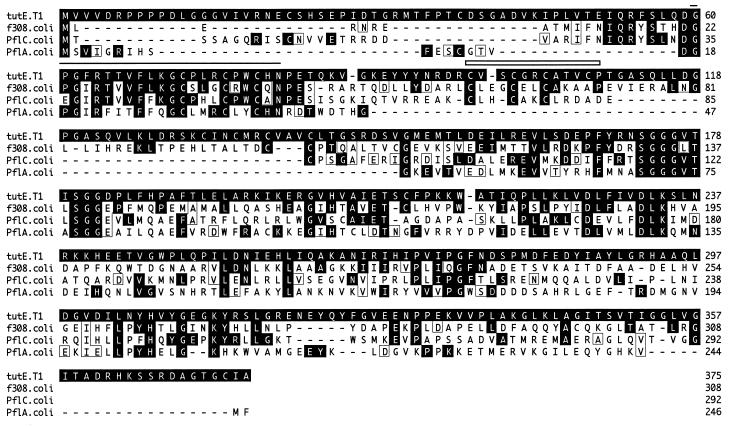
FIG. 3.
Comparison of the predicted amino acid sequence of the TutE protein with the predicted sequences of the f308 protein of E. coli (7), the PflC protein of E. coli (6), and the PflA protein of E. coli (31). The region defined as a radical-activating region is indicated by a line (positions 60 to 81 of TutE). The region defined as a 4Fe-4S binding domain typical of ferredoxins is indicated by an open box (positions 98 to 109 of TutE). This motif was identified only in TutE. Amino acids identical to the tutE translation are shaded, and conserved amino acids are boxed. Dashes indicate gaps introduced by the computer program to maximize the alignment score.

