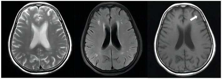
Figure 4.
Unenhanced brain MRI scan on April 19, 2022(D + 74): Multiple infarcts and ischemic foci were seen bilaterally in the periventricular and frontoparietal white matter; there were abnormally enhancing lesions in the right ambient cistern, left sylvian cistern and adjacent brain parenchyma, which was considered a diagnosis of TBM, especially by referring to her medical history; also there was mild brain atrophy.

