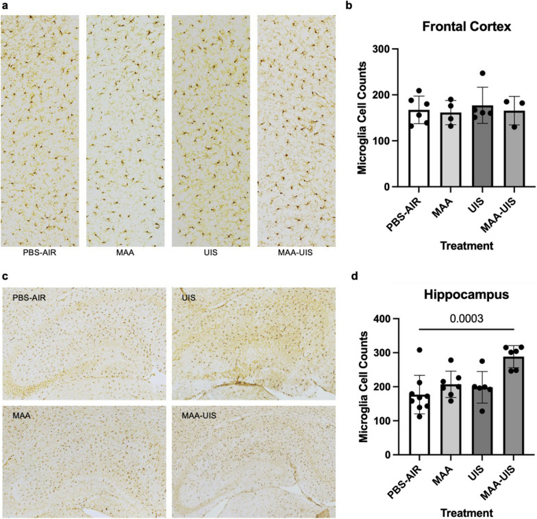
Fig. 4.
Microglia density in the hippocampus and frontal cortex of MAA, UIS, and MAA-UIS of P15 offspring. a and c Staining for IBA-1 with diaminobenzidine (DAB) was used to label microglia in coronal sections of p15 mice in the (a) frontal cortex and (c) hippocampus. b and d Quantification of microglial density in the (b) frontal cortex and (d) hippocampus. Statistical significance determined via one-way ANOVA. In the frontal cortex, n = 6 (PBS-AIR), n = 4 (MAA), n = 5 (UIS), n = 3 (MAA-UIS). In the hippocampus, n = 9 (PBS-AIR), n = 7 (MAA), n = 6 (UIS), n = 6 (MAA-UIS)

