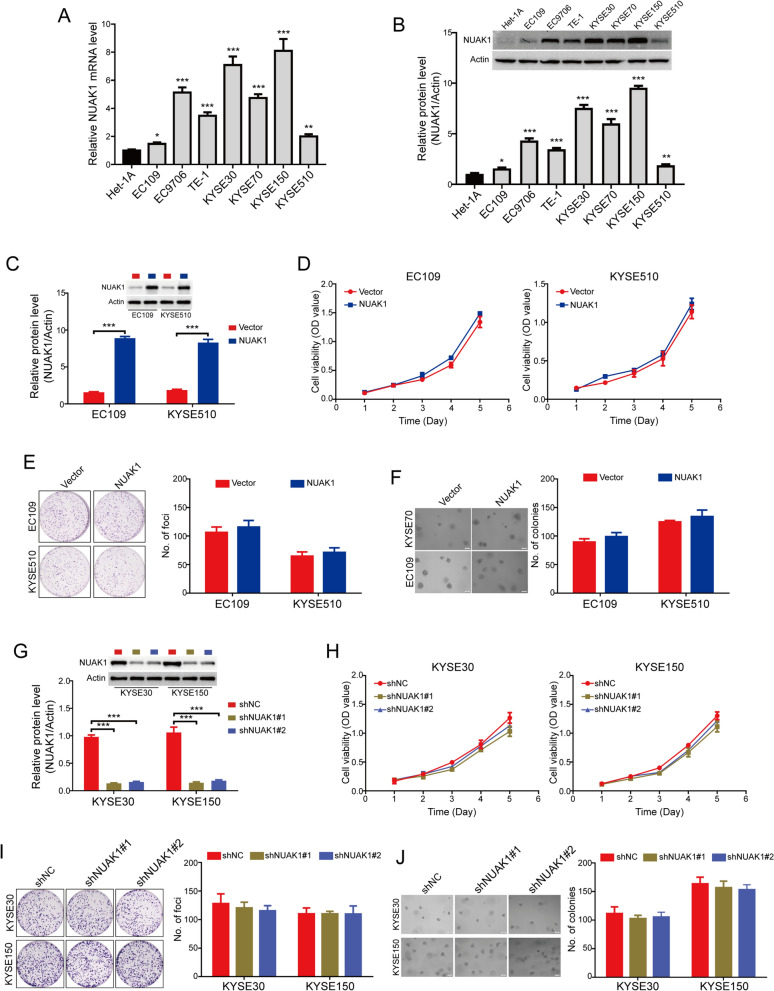
Fig. 2.
NUAK1 does not influence the tumor cell growth in ESCC cells. A, B The expression of NUAK1 in ESCC cells at mRNA and protein levels were detected by qRT-PCR (A) and Western blotting (B), respectively. P value was calculated by one-way ANOVA with post hoc intergroup comparison by the Tukey’s test. C Western blot analysis was performed to detect the level of NUAK1 in EC109 and KYSE510 cells stably expressing NUAK1. Quantification of indicated protein levels of n = 3 independent biological experiments (low panel). D–F The effects of NUAK1 overexpression on ESCC cell growth were assessed by MTT assay (D), foci formation assay (E) and soft agar assay (F). P value was calculated by Student’s t-test (for C–F). G KYSE30 and KYSE150 cells stably expressing shNC or NUAK1 shRNA as indicated were analyzed by Western blotting. Quantification of indicated protein levels of n = 3 independent biological experiments (low panel). H–J The effects of NUAK1 knockdown on ESCC cell growth were assessed by MTT assay (H), foci formation assay (I) and soft agar assay (J). P value was calculated by one-way ANOVA with post hoc intergroup comparison by the Tukey’s test (for G–J). Error bars denote mean ± SD. *P < 0.05; **P < 0.01; ***P < 0.001

