
Figure 2.
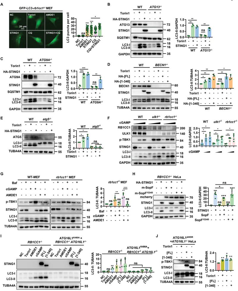
STING1 can significantly induce non canonical autophagy in ULK1- and BECN1-deficient cells. (a) rb1cc1−/− MEFs stably expressing GFP-LC3 with or without transfected of HA-STING1 plasmid were treated with indicated chemicals (AMDE-1, 10 μM; CQ, 20 μM; torin1, 2 μM) for 6 h. Quantification of LC3 puncta per cell was shown on the right side. 50 randomly-selected cells per condition were counted. (b-c) Western blotting of cell lysates from WT-HeLa, ATG13-/- HeLa, and ATG9A-/- HeLa cells were treated with torin1 (2 μM) for 6 h or transfected with HA-STING1 plasmid for 24 h. (d) WT and BECN1−/− HEK-293T cells were treated with torin1 (2 μM) for 6 h or transfected with HA-STING1[FL] or the C-terminal truncation (STING1 [1-340]) plasmids for 24 h. Cell lysates were subjected to western blotting analysis with the indicated antibodies. (e) WT and atg5-/- MEF cells were treated with torin1 (2 μM) for 6 h or transfected with HA-STING1 for 24 h. Cell lysates were subjected to western blotting analysis with the indicated antibodies. (f) WT-MEFs, rb1cc1-/- MEFs and ulk1-/- MEFs were treated with increasing amounts of cGAMP (0 µM, 1 µM and 2 µM respectively) for 3 h, followed by western blotting assays of LC3-ΙΙ conversion. (g) WT and rb1cc1-/- MEF cells were treated with indicated chemicals (AMDE-1, 10 μM; bafilomycin A1 (Baf), 0.5 μM; cGAMP, 1 μM) for 6 h. Immunoblotting was then performed to analyze the LC3-ΙΙ conversion. (h) Western blotting of cell lysates from RB1CC1−/− HeLa cells lacking endogenous STING1 expressing were transfected with HA-STING1 or mcherry-SopF and its mutant mcherry-SopFY224D plasmids for 24 h, and then assessed for LC3-ΙΙ conversion in the right panel. (i) RB1CC1−/− HeLa cells and RB1CC1 and ATG16L1 double knockout cells stably expressing ATG16L1K490A were treated with indicated chemicals (AMDE-1, 10 μM; cGAMP, 1 μM; Torin1, 2 μM) for 6 h or transiently transfected with HA-STING1[FL] or the C-terminal truncation (STING1 [1-340]) plasmids for 24 h, and then assessed for LC3-ΙΙ conversion in the right panel. (J) Restoring the ATG16L1ΔWDR domain by stably expressing on ATG16L1−/− HeLa cells treated with torin1 (2 µM) for 6 h or transiently transfected with HA-STING1[FL] or the C-terminal truncation (STING1 [1-340]) plasmids for 24 h. Cell lysates were subjected to western blotting analysis with the indicated antibodies.
*P<0.05, **P<0.01, ***P<0.001, ns, not significant.