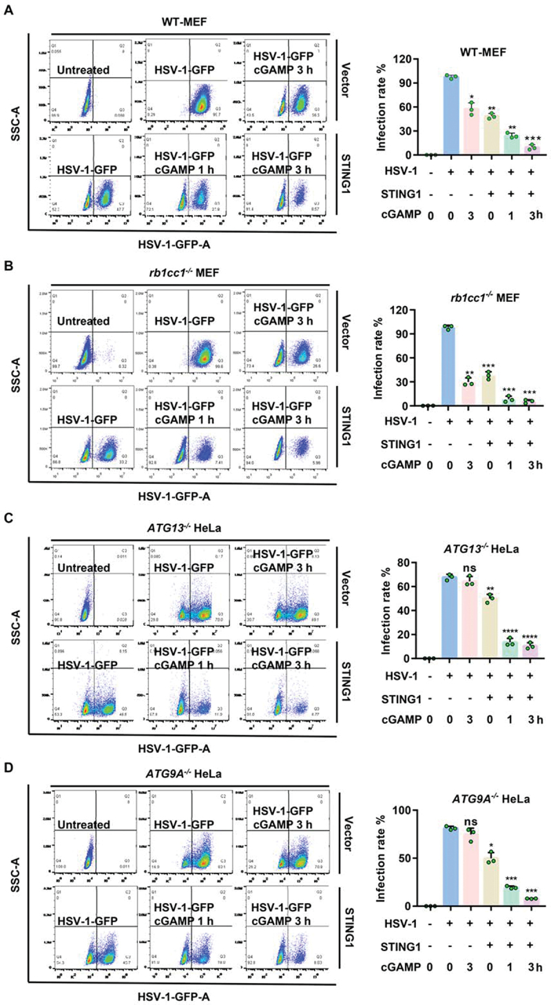
Figure 3.

STING1 show obvious resistance to HSV-1 virus in both WT and autophagy gene deficient cells. (A-D) WT (A) or rb1cc1−/− MEF (B) cells containing endogenous STING1 expressing [11] and ATG13−/− HeLa (C) and ATG9A−/− HeLa (D) cells lacking endogenous STING1 expression were transfected with HA-STING plasmids. After 24 h incubation, cells were then infected with HSV-1-GFP virus at a multiplicity of infection (MOI) of 5 for 6 h, with or without 1 μM of cGAMP stimulated for the indicated time. GFP-positive cells were analyzed by flow cytometry. Quantification charts of HSV-1-GFP virus infection rate in (A-D) were shown in the right panels. Data analysis was performed using FlowJo software and presented as mean±SEM from 3 individual experiments.
*P<0.05, **P<0.01, ***P<0.001 vs with positive control based on HSV-1-GFP infection alone, ns, not significant.
