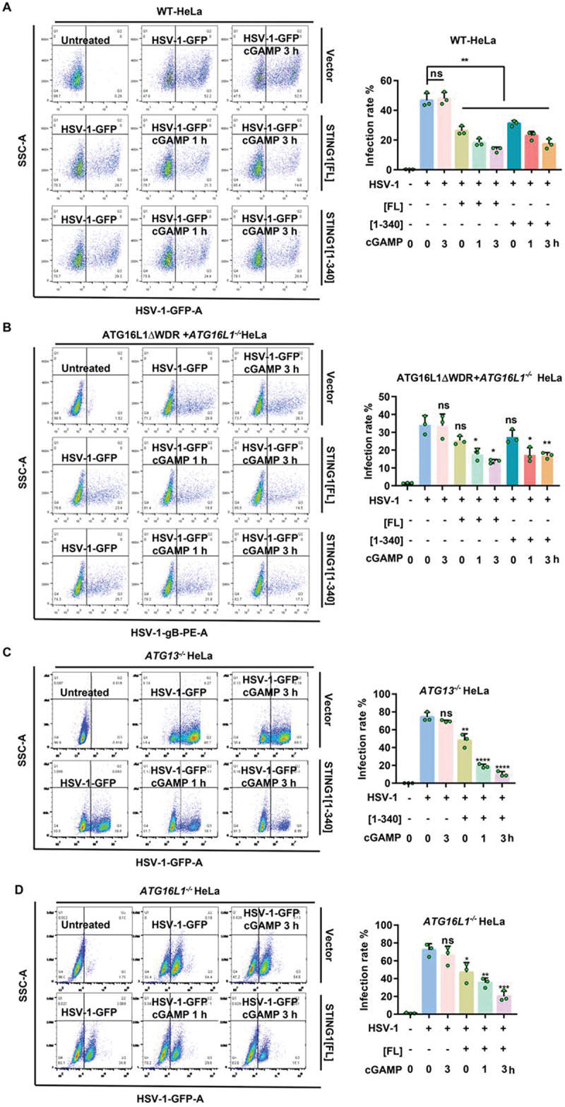
Figure 4.

STING1 can inhibit HSV-1 through noncanonical autophagy pathway alone, canonical autophagy pathway alone, or immunity function alone. (A) WT HeLa cells which lacking endogenous STING1 expressing were transfected with STING1 truncated mutants STING1 [1-340] plasmids or full-length STING1[FL] plasmid. After 24 h expression, cells were infected with HSV-1-GFP virus at a MOI of 2.5 for 6 h, with or without 1 μM of cGAMP stimulated for the indicated time. GFP-positive cells were analysis by flow cytometry. Quantification of HSV-1-GFP virus infection rate is shown in the right panel. (B) After STING1[FL] or STING1 [1-340] plasmids were transfected into NCA-deficient ATG16L1ΔWDR-GFP HeLa cells for 24 h, HSV-1-GFP virus were then infected for 6 h, with or without 1 μM of cGAMP stimulated for the indicated time. PE flow detection channel was used to detect HSV-1-GFP virus coat gB protein to avoid the interference of GFP in the basal of the ATG16L1ΔWDR-GFP ATG16L1-/-HeLa cells. PE positive cells were analysis by flow cytometry. (C-D) ATG13-/- HeLa cells which have canonical autophagy dysfunction (C) and ATG16L1-/- HeLa cells which have autophagy dysfunction (D) were transfected with STING1 truncated mutants STING1 [1-340] (C) or full length STING1 (D). After 24 h expression, cells were then infected with HSV-1-GFP virus at a MOI of 5 for 6 h, with or without 1 μM of cGAMP stimulated for the indicated time. GFP-positive cells were analyzed by flow cytometry. Quantification charts of HSV-1-GFP virus infection rate were shown in the right panels. Data analysis was performed using FlowJo software and presented as mean±SEM from 3 individual experiments.
*P<0.05, **P<0.01, ***P<0.001 vs with positive control based on HSV-1-GFP infection alone, ns, not significant.
