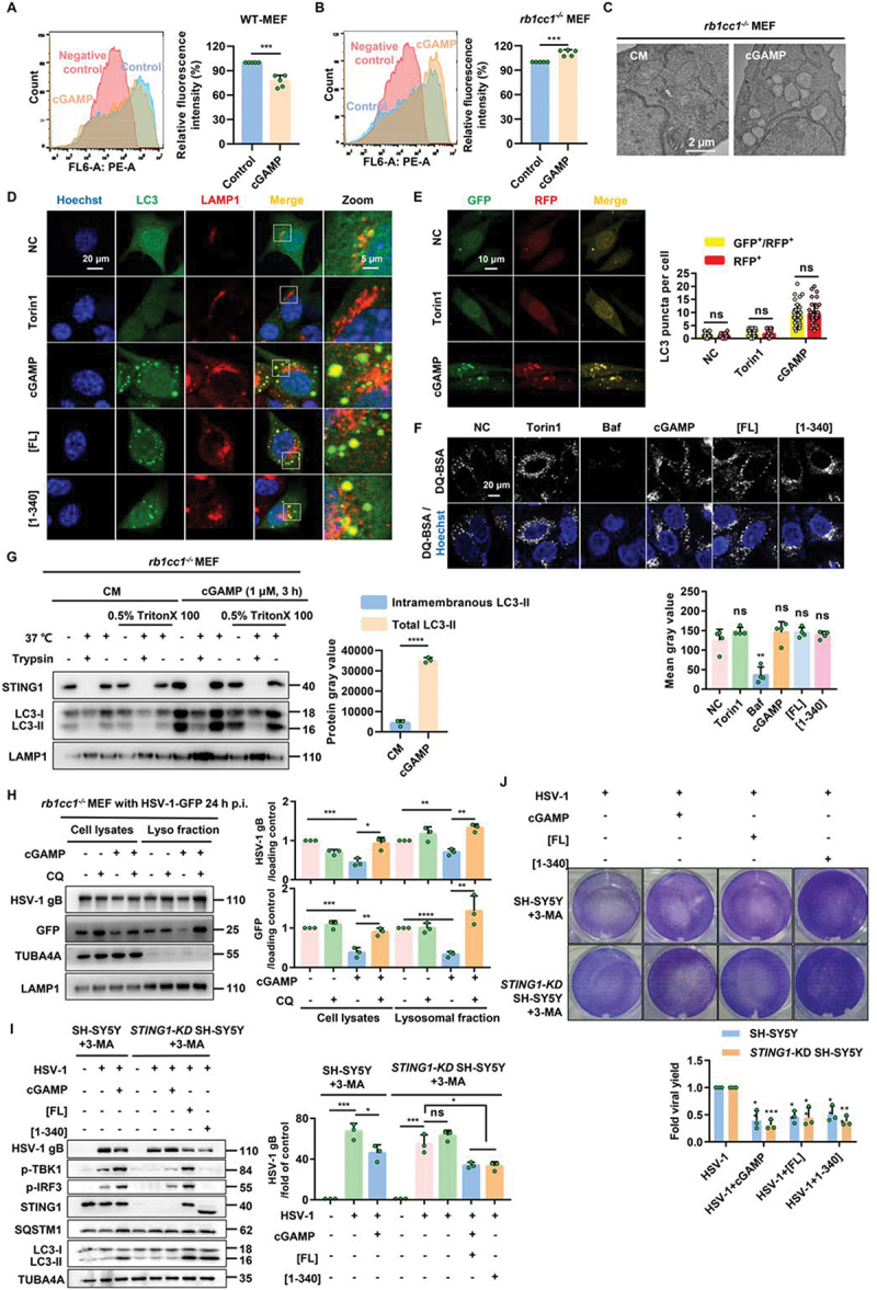
Figure 7.

Non canonical autophagy induced by STING1 can degrade the virus through lysosomes.(A-B) Flow cytometry was performed to analyze the fluorescence intensity within the WT MEF (A) and rb1cc1-/- MEF (B) cells, and the histograms represent the relative average fluorescence intensity of 10,000 cells from different samples. Negative control: cells without AHA labeling but were subjected to click reaction. Data for the relative signal intensity were expressed as the ratio of 1 μM of cGAMP treated for 6 h cells to the untreated control cells. (C) Representative images of transmission electronic microscope of rb1cc1-/- MEF cells stimulated with 1 μM of cGAMP for 6 h. (D) Colocalization of NCA autophagosomes and lysosomes induced by STING1. rb1cc1-/- MEF cells stably expressing GFP-LC3 were treated with indicated chemicals (torin1, 2 μM; cGAMP, 1 μM) for 6 h, or transfected with STING1 plasmids (STING1[FL] or STING1 [1-340]) for 24 h, followed by immunostaining of LAMP1 (red). Scale bar: 20 µm. (E) rb1cc1-/- MEF cells which containing endogenous STING1 expressing were transiently transfected with RFP-GFP-LC3 plasmid. After 24 h expression, cells were then harvested and re-seeded on confocal culture dish. Cells were treated with indicated chemicals (torin1, 2 μM; cGAMP, 1 μM) for 6 h, followed by fixation. The colocalization of GFP and RFP puncta was examined and quantified. Scale bar: 10 µm. At least 50 cells were counted from each group. (F) Representative single-plane confocal micrographs of WT HeLa cells incubated with DQ-Red BSA were starved for 2 h by EBSS in the absence or presence of 500 nM Baf, 2 μM torin1, 1 μM cGAMP or transfected with STING1 plasmids (STING1[FL] or STING1 [1-340]) for 24 h. The cell nucleus is stained using DAPI (blue). Scale bar: 20 µm. And then assessed for average fluorescence value of DQ-BSA in the right panel. At least 50 cells were counted from each group. (G) Protease protection assay of homogenates from rb1cc1−/− MEF cells treated with 1 μM of cGAMP for 3 h. Diagrams show subcellular location of LC3-II and endogenous STING1 detected by western blotting. (H) Detect the virus content in lysosomes. Rb1cc1-/- MEF cells which contain endogenous STING1 were infected with HSV-1 virus at MOI = 5. After 24-h infection, cells were harvested and re-seeded on culture dish. Cells were then treated with indicated chemicals (CQ, 40 μM; cGAMP, 1 μM) for 6 h. Lysosomal fraction was collected, followed by immunoblotting with indicated antibodies. Representative data were shown from three independent experiments; n = 3.
*P<0.05, **P<0.01, ***P<0.001, ns, not significant.
