
Figure 7.
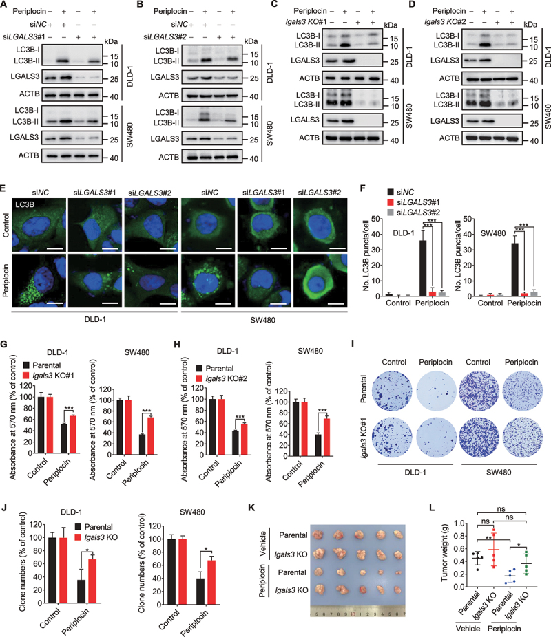
Periplocin induces lethal lysophagy by upregulating LGALS3 in CRC cells. (A and B) Immunoblotting analysis of LC3B turnover in cells transfected with siNC or siLGALS3 followed by 0.50 μM periplocin treatment for 24 h. (C and D) Immunoblotting analysis of LC3B turnover in parental or lgals3 KO cells followed by 0.50 μM periplocin treatment for 24 h. (E and F) Representative images (E) and quantitative analysis (F) for immunofluorescent staining of endogenous LC3B puncta in cells transfected with siNC or siLGALS3 followed by 0.50 μM periplocin treatment for 24 h. Scale bar: 10 μm. (G and H) MTT assay of CRC cells with or without LGALS3 knockout (G, lgals3 KO#1; H, lgals3 KO#2) in response to 0.50 μM periplocin treatment for 24 h. (I) Colony formation assay of parental or lgals3 KO cells treated with or without 0.50 μM periplocin for 24 h. (J) Quantification of clone numbers in (I). (K and L) DLD-1 parental or lgals3 KO cells were subcutaneously inoculated into nude mice. Mice were injected with vehicle or periplocin (15 mg/kg/day) for two weeks. Image (K) and weight (L) of tumor xenografts were shown. Results in A-J are representative of three independent experiments. Data are presented as mean ± SD. *P < 0.05, **P < 0.01, ***P < 0.001. ns, non-significant.