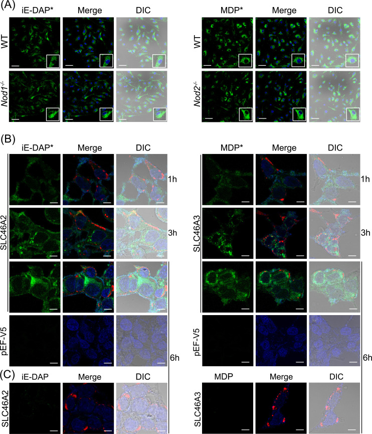
Figure 7.
Internalization of iE-DAP-Alk and MDP-Alk in BMDMs and HCT116 cells. (A) Using click-chemistry with AZDye-488 (green), the intracellular distribution of iE-DAP-Alk or MDP-Alk, independent of Nod1 or Nod2, was observed by confocal fluorescent microscopy in IFNγ-primed bone marrow-derived macrophages at 6hr. Nuclei stained with Hoechst (blue) (B) HCT116 cells transiently expressing mouse Slc46a2 or Slc46a3 similarly showed intracellular iE-DAP-Alk or MDP-Alk uptake (green) that increased with time from 1 to 6 h, whereas empty vector-transfected cells failed to import these NOD1/2 agonists. (C) Additionally, cells treated with native iE-DAP and MDP (without Alk-modification) did not react with AZDye-488. Throughout Panels B and C, the red stain is V5 tagged Slc46a2 or Slc46a3. Nuclei stained with Hoechst (blue). Images in panels are representative of three independent experiments. The scale bar in panel A is 50 µM and in panels B and C is 10 µM.

