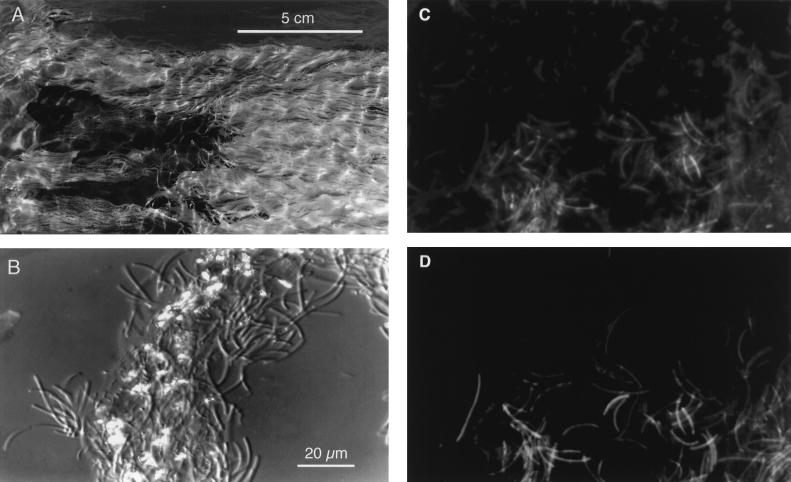
FIG. 1.
Photographs of a sulfur-turf microbial mat. (A) Ruffled fur or turf-like appearance of a mat in a shallow hot spring stream; (B) Nomarski interference contrast micrograph of the sulfur-turf mat consisting of bundles of large sausage-shaped bacteria and glittering elemental sulfur particles; (C) epifluorescence microscopic image of the sulfur-turf mat stained with DAPI; (D) image of fluorescently labeled probe hybridized to large sausage-shaped cells in the same microscopic field shown in panel C. The microscopic images were obtained with a charge-coupled device camera (model C5910; Hamamatsu Photonics K.K., Hamamatsu, Japan).

