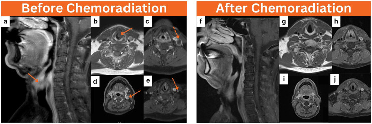
Figure 1.
Baseline neck MRI was performed before chemoradiation, coronal T1 neck MRI (a) and axial T1 images (b) revealed a left transglottic laryngeal mass infiltrating the left paraglottic space and measuring about 2.5 cm in maximum dimension (arrows). In addition, axial T1 images of the neck (c–e) showed few concurrent ipsilateral enlarged level II–IV cervical lymph nodes, the majority of which demonstrate central necrosis (dotted arrows). At the end of therapy, coronal T1 neck MRI (f) and axial T1 neck images (g–j) concluded resolution of the previous locoregional disease.

