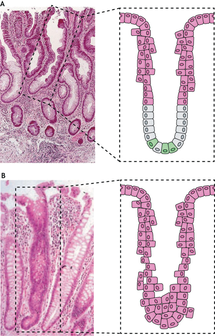
Fig. 1.

Early features of colon crypt morphogenesis in human adenomas. (A) Top-down morphogenesis. H&E-stained section of a typical small adenomatous polyp in the human colon. The highlighted structure shows dysplastic epithelium at the top of a crypt that is contiguous with the histologically normal epithelium at the base of the crypt. Notice the abrupt transition between dysplastic cells (pink) and the normal-appearing crypt stem cells and differentiating epithelial cells near the crypt mid-point (green and grey). The photomicrograph was reproduced with permission from Shih et al. (2001a). (B) Bottom-up morphogenesis. H&E-stained colonic section from an individual with familial adenomatous polyposis showing a monocryptal adenoma, in which all cells are dysplastic (pink). Dysplastic stem cells that are located at the crypt base always drive the dysplastic morphology for an entire crypt. The photomicrograph was reproduced with permission from (Preston et al., 2003).
