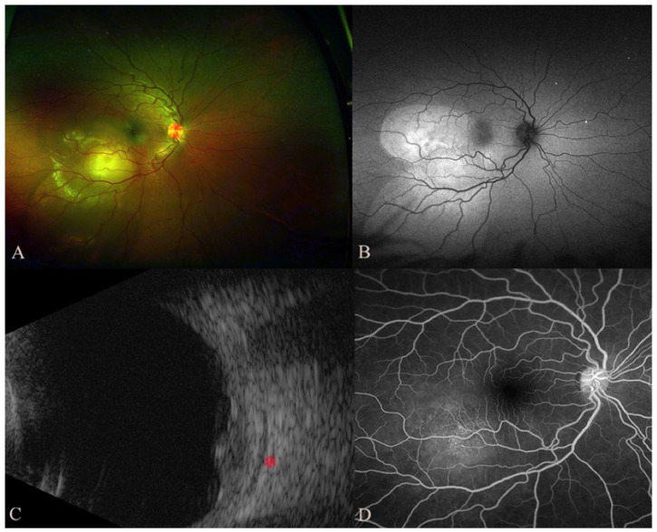
Figure 1.
On initial presentation, the color fundus photograph (A) shows an amelanotic choroidal elevation with surrounding subretinal fluid that was highlighted on the autofluorescence image (B) as an area of hyperautofluorescence that was most pronounced over the lesion with areas of inferior, gravity-dependent guttering. (C) Ultrasonography shows a choroidal lesion with moderate internal reflectivity and associated fluid in Tenon space (red asterisk). (D) Fluorescein angiogram from the late arteriovenous phase shows multiple pinpoint spots of leakage overlying the lesion.

