
Fig 8. Simulating fusion of multicellular, homotypic spheroids.
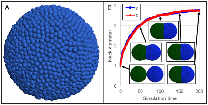
A: Spheroids of 12.5k cells each were individually pre-assembled, as in typical bioprinting practice. B: Two spheroids (green and blue) placed in close proximity fuse over time, as measured by the neck diameter along the y (blue circles) and z (red triangles) directions, which grows over time. The neck diameter along a direction is measured as the largest distance along the direction between any two particles at the mid-plane. Insets show the simulation at times 1, 50, 100, 150 and 200.