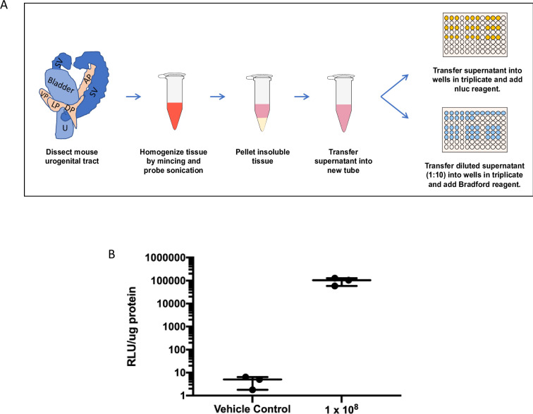
Fig 3. Quantifying parasite burden using nanoluciferase-expressing Tv.
(A) Schematic of mouse MUT tissue sample preparation and quantification of Tv parasite burden. Excised mouse MUT tissue is finely minced and suspended in prostate lysis buffer (2 mL/mg tissue). MUT tissue then undergoes additional homogenization via probe sonication to ensure complete tissue dissociation. Insoluble material is pelleted via centrifugation and the supernatant is transferred to a new tube to further quantify luciferase activity and protein concentration using the Nano-Glo luciferase and Bradford assays, respectively. Detailed description found in materials and methods. (B) Mice were inoculated with 108 parasites (n = 3) or RPMI vehicle control (n = 3) and promptly euthanized. Samples were processed and analyzed by Nluc and Bradford assays to quantify parasite load. Data is shown as average luminescence signal/μg protein of the sample.

