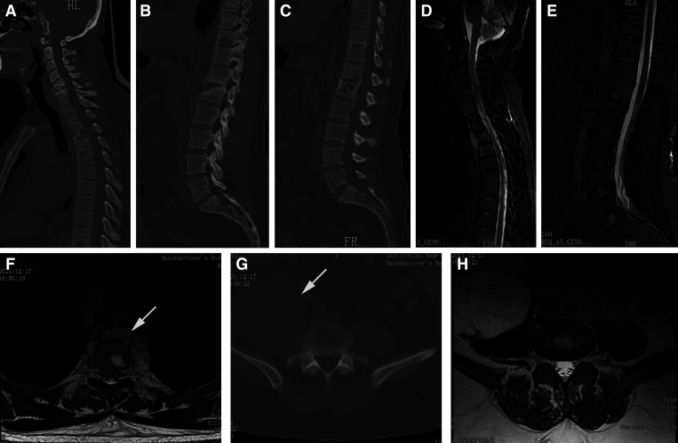
Figure 1.
Preoperative CT and MRI scans. (A–C) Sagittal CT images indicate bony destruction in the C5, C6, T3, T10, T12, and L1 vertebrae. (D and E) Preoperative sagittal MRI indicates high signal intensity in multiple vertebrae and a fusiform paraspinal abscess in front of the T1–T4 vertebrae. (F) Axial MRI scan at the T4 vertebra level indicate a paravertebral abscess (white arrow). (G and H) Axial CT and MRI scans at the L4/5 intervertebral level indicate a massive psoas abscess (white arrow). CT = computed tomography; MRI = magnetic resonance imaging.

