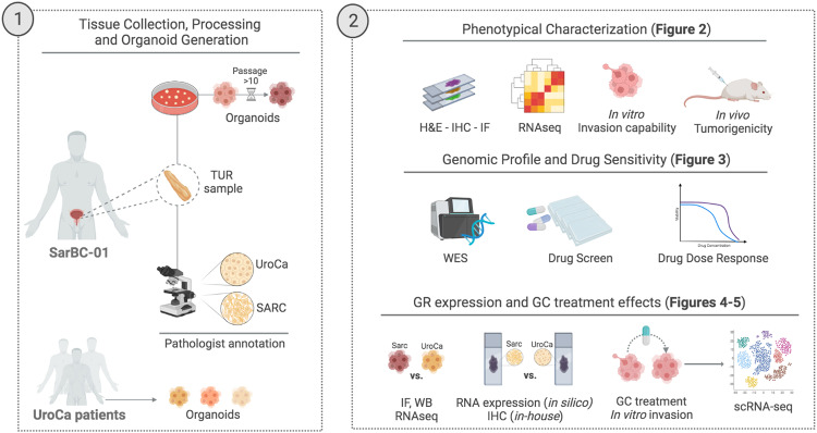
Fig. 1. Cartoon depicting the distinct morphological components of the patients’ tumor and the experimental workflow used to generate organoids (1) and subsequent analyses (2).
UroCa urothelial carcinoma, SARC sarcomatoid urothelial bladder cancer, TUR transurethral resection, H&E hematoxylin and eosin, IHC immunohistochemistry, IF immunofluorescence, WES whole exome sequencing, WB western blot, GR glucocorticoid receptor, GC glucocorticoids. Created with BioRender.com.

