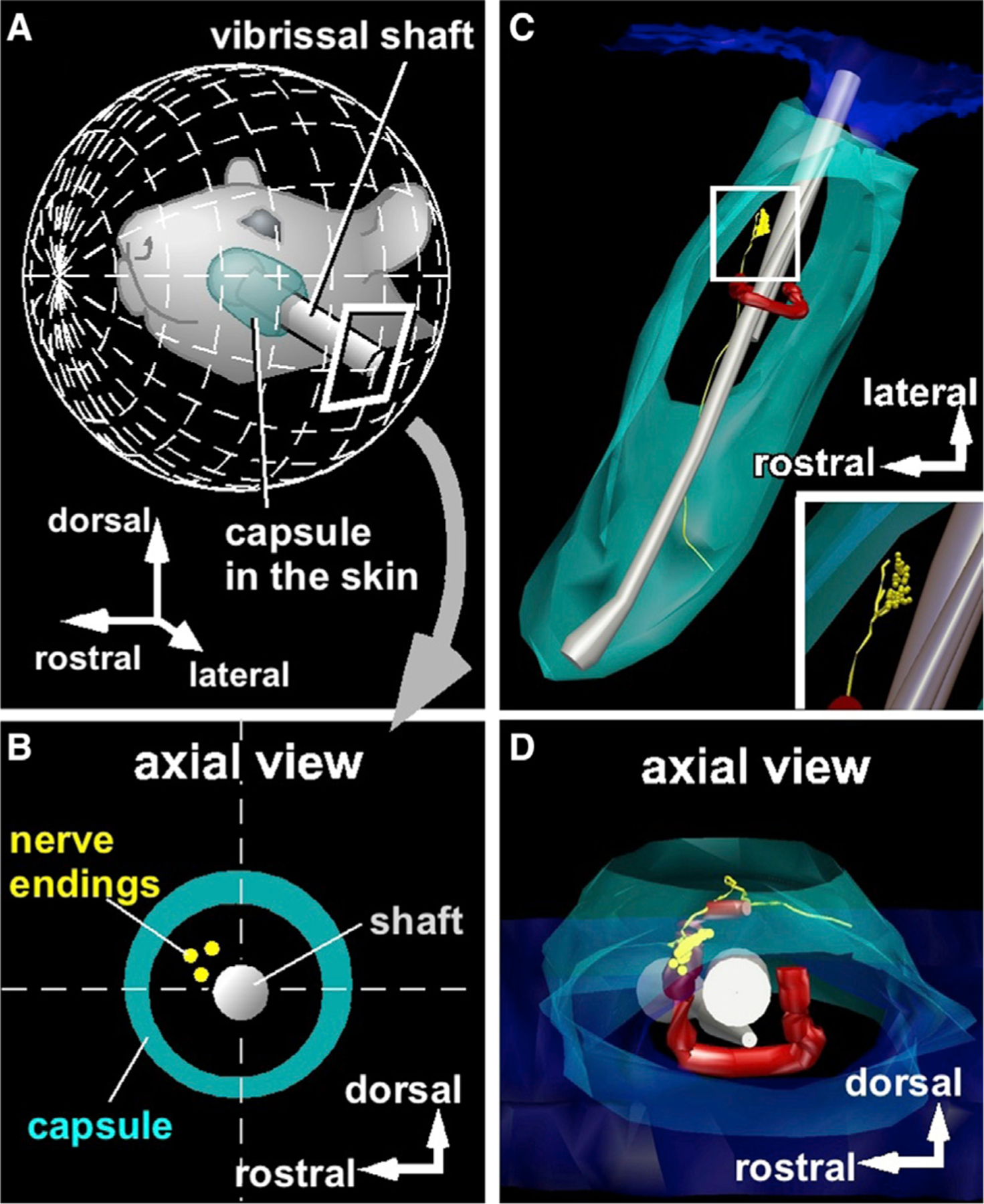
Figure 3. Identifying the 3D Location of the Mechanoreceptor in the Follicle.

(A) Vibrissa orientation relative to mystacial pad.
(B) Locations of peripheral endings were projected into a plane normal to the vibrissal length.
(C) Representative 3D reconstruction of a vibrissal follicle. Peripheral endings are yellow.
(D) Reconstructed data are observed from a direction along the vibrissal length.
See also Figures S2 and S3.
