
Figure 6. Ultrastructure of RS-Merkel and Lanceolate Endings.
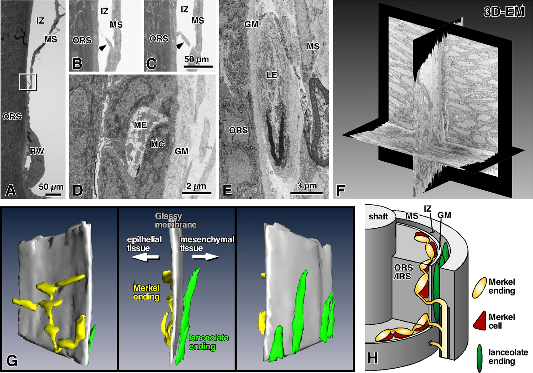
GM, glassy membrane (basement membrane); IRS, inner root sheath; IZ, intermediary zone; LE, lanceolate ending; MC, Merkel cell; ME, Merkel ending; MS, mesenchymal sheath; ORS, outer root sheath; 3D-EM, three-dimensional electron microscopy.
(A) Semi-thin section parallel to the axis of the vibrissa.
(B and C) Magnified views of the rectangle in (A) obtained from two sequential semi-thin sections. Arrowheads indicate an axon branch piercing the glassy membrane to enter the ORS.
(D)RS-Merkel endings are distributed in the most lateral layer of the ORS.
(E) Ultrastructure of a lanceolate ending.
(F) Three orthogonal planes of stack data obtained with a scanning electron microscopic system.
(G) Sequential EM images of nerve endings at the ring sinus level were reconstructed in 3D (gray, glassy membrane; yellow, Merkel ending; green, lanceolate ending). RS-Merkel endings are located in the epithelial sheath although lanceolate endings are localized in the loose space between the glassy membrane and the mesenchymal tissue.
(H) Schematic representation of Merkel and lanceolate ending innervation at the level of the ring sinus.