
Figure 2.
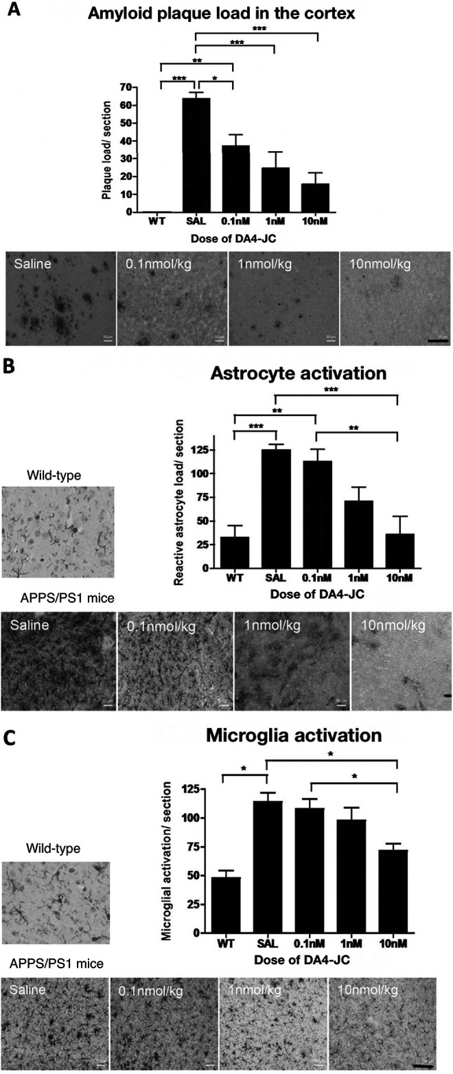
DA4-JC4 dose-response study in the APP/PS1 mouse model of AD. A: Immunohistochemical analysis of amyloid plaque load in the cortex of 9-month old APP/PS1 mice. The 4 different treatments given to the APP/PS1 mice, are shown in the images above, presenting a reduced number of amyloid plaques in the cortex (scale bar 100 µm). Data shown as mean ± S.E.M. (One-way ANOVA, Tukey’s Multiple Comparison post-hoc test, *=p < 0.05, **=p < 0.01, ***=p < 0.001). B: Immunohistochemical analysis of the number of reactive astrocytes in the cortex of 9-month old APP/PS1 mice. DA4-JC treatment reduced the number of activated astrocytes in the brain. The 5 images are illustrating the different treatments given to the APP/PS1 mice, showing the reduction in the number of reactive astrocytes per section within the cortex (scale bar 100 µm). Data shown as mean ± S.E.M. (*=p < 0.05, **=p < 0.01, ***=p < 0.001). C: Immunohistochemical analysis of the number of activated microglia in the cortex of 9-month old APP/PS1 mice. DA4-JC treatment reduced the number of activated microglia in the brain. The 5 images are illustrating the different treatments given to the wild-type and APP/PS1 mice, showing the reduction in activated microglia within the cortex (scale bar 100 µm). Data shown as mean ± S.E.M. (*=p < 0.05, **=p < 0.01, ***=p < 0.001).