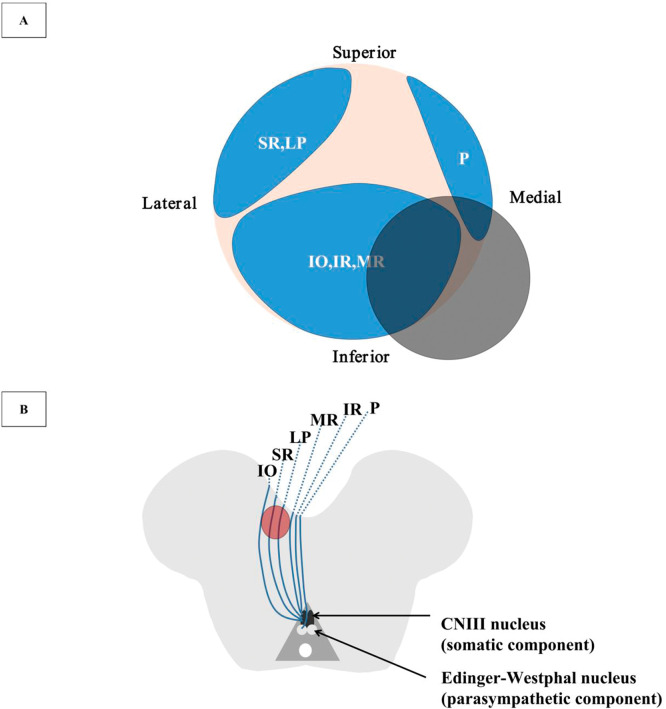
Figure 2. Illustration of the CNIII Microanatomy in the Cistern Space and the Diagram of the CNIII Nerve Fascicular Topography in the Midbrain (Not to Scale).
(A) Illustration of the CNIII microanatomy. The gray shaded area demonstrates the region of the neurovascular conflict in our patient showing the possibly involved CNIII fascicles, which is not in accordance with the symptomatology. (B) Diagram of the topographic fascicular arrangement of the CNIII nerve. The gray shaded area shows the midbrain lesion in our patient. CNIII = third cranial nerve; IO = inferior oblique; IR = inferior rectus; LP = levator palpebrae; MR = medial rectus; P = pupillary fibers; SR = superior rectus.

