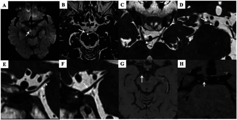
Figure 3. Neuroimaging.
(A) Restricted diffusion involving the medial aspect of the right cerebral peduncle. (B–F) Axial (B, C), coronal (D, showing right CNIII nerve), and sagittal (E, right CNIII nerve; F, left CNIII nerve) 3D-CISS MRI showing the right SCA (arrowhead) indenting the inferior and medial part of the CNIII nerve (arrow) in the cistern space, resulting in nerve extension and deviation. HRMR-VWI showed stenosis and a plaque in the origin of right PCA (G–H, arrow). 3D-CISS = 3-dimensional constructive interference in steady state; CNIII = third cranial nerve; HRMR-VWI = high-resolution magnetic resonance vessel wall imaging; PCA = posterior cerebral artery.

