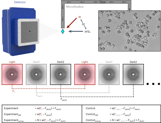
Fig. 1. Schematic of T-jump TRX experiment.
Lysozyme crystals were delivered to the pump–probe interaction region via a microfluidic jet. Light and dark images were collected in an interleaved manner, with light images defined as those where the crystal was pumped with an IR laser at a defined time delay (∆t) before being probed by the XFEL. Dark images were collected with the IR pump shutter closed and an XFEL probe. These images were combined in post-processing to create a set of structure factors for each time delay (Ft) and as well as two corresponding sets of dark structure factors (Fdark1 and Fdark2). Experiments and matching controls as defined above and in the text were analysed to identify time-dependent structural changes.

