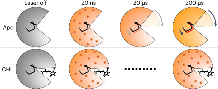
Fig. 6. Schema of time-resolved conformational changes in lysozyme following T-jump.

The cartoon highlights closure of the active site cleft upon chitobiose (CHI) binding, with subsequent representations of time-resolved structural changes following T-jump. At short pump–probe time delays (20 ns) atomic vibrations (shown as red dots) are present in both the apo and inhibitor-bound structures. These vibrations persist in the inhibitor-bound structure but dissipate into more complex motions in the apo structure, including the bending of a helix that lies at the hinge point of the lysozyme molecule.
