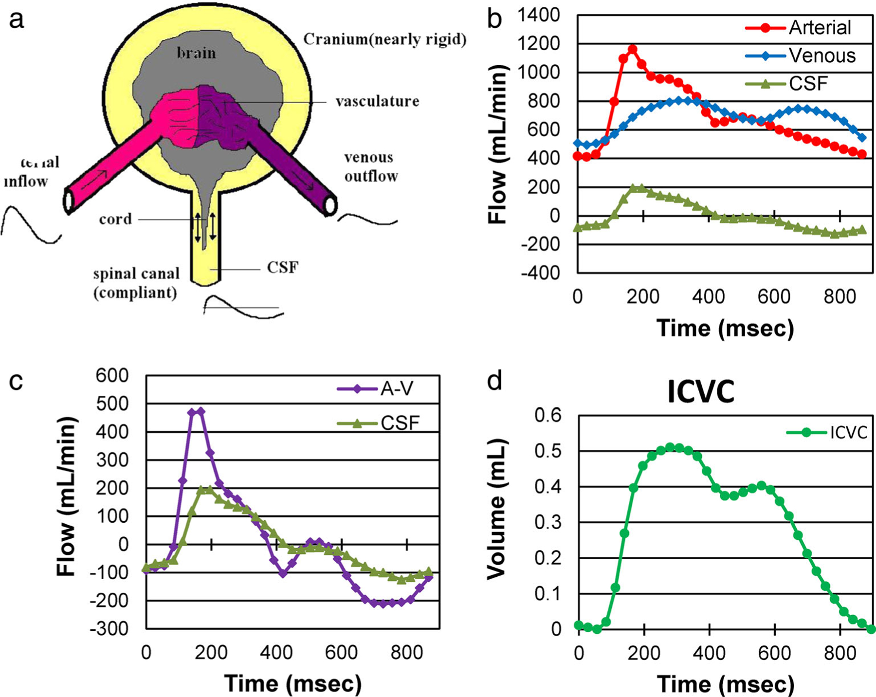
FIGURE 5:

(a) A simplified model of the CS system. (b) Volumetric flow rate waveforms of the arterial inflow, venous outflow, and the CS CSF flow. (c) The cs CSF flow waveform plotted with respect to the arterial minus venous (A-V) flow waveform. The fact that these two waveforms are not identical implies that the ICV is not constant and thus, the cranium has compliance. The CSF waveform follows the pattern of the net transcranial blood flow suggesting that the A-V flow drives the CS CSF pulsation. (d) The intracranial volume change during the cardiac cycle obtained by time integration of the next transcranial flow.48
