Figure 3.
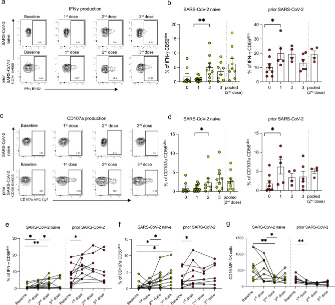
Antibody-dependent NK cell responses following SARS-CoV-2 vaccination in PLWH. (a) Representative flow cytometric plots and (b) cross-sectional analysis of the percentage of IFN-γ+ CD56dim NK cells from PLWH, with or without prior SARS-CoV-2 infection, at baseline (pre-vaccine) and following each dose of vaccine. (SARS-CoV-2 naïve: n = 14 baseline, n = 9 first dose, n = 12 s dose, n = 8 third dose; prior SARS-CoV-2 n = 12 baseline, n = 7 first dose, n = 5 s dose, n = 7 third dose). (c) Representative flow cytometric plots and (d) cross-sectional analysis of frequencies of CD107a+ CD56dim NK cells. Longitudinal analysis of (e) IFN-γ+ CD56dim NK cells, (f) CD107a+CD56dim NK cells, and (g) CD16 MFI in the two study groups. Significance determined by two-tailed Mann–Whitney U test [(b), (d)], or two-tailed Wilcoxon-signed rank test [(e), (f), (g)]; *p < 0.05, **p < 0.01. Data are represented as geometric mean ± SEM [(b), (d)]. See also Supplementary Fig. 3.

