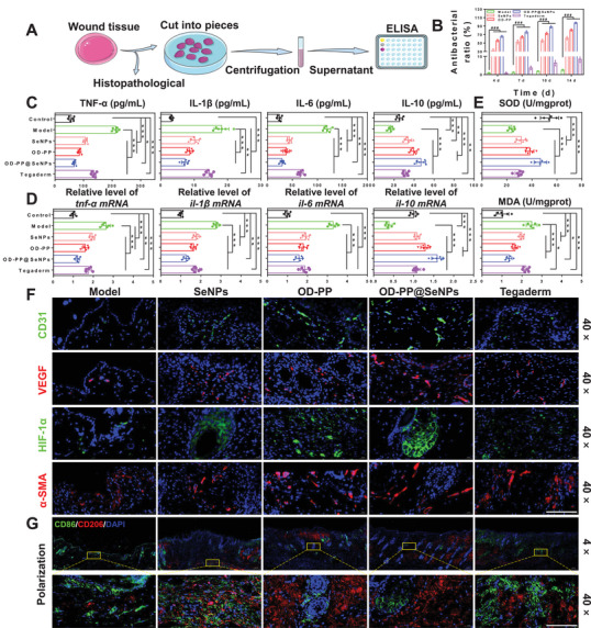
Figure 6.

In vivo antibacterial, anti‐inflammatory, and vascularization capacity of the OD‐PP@SeNPs hydrogel. A) A schematic diagram of the wound tissue detection process. B) Antibacterial effect of different preparations in vivo (n = 3). C) Expression levels of TNF‐α, IL‐1β, IL‐6, and IL‐10 after treatment with the different preparations at wound tissue (n = 6). D) Gene expression levels of tnf‐α, il‐1β, il‐6, and il‐10 after treatment with the different preparations at wound tissue (n = 6). E) MDA and SOD levels at the wound site (n = 6). F) Immunofluorescence staining images of CD31 (green), VEGF (red), HIF‐1α (green), α‐SMA (red), and DAPI (blue) at the wound tissue with different treatments at 14 d. Scale bar = 200 µm. G) Immunofluorescence staining of macrophage phenotype at the wound tissue with different treatments at 14 d. CD86 (green), CD206 (red), and DAPI (blue). Scale bar = 200 µm.
