
Fig. 1 |. Endogenous plasticity in sub-acute stroke.
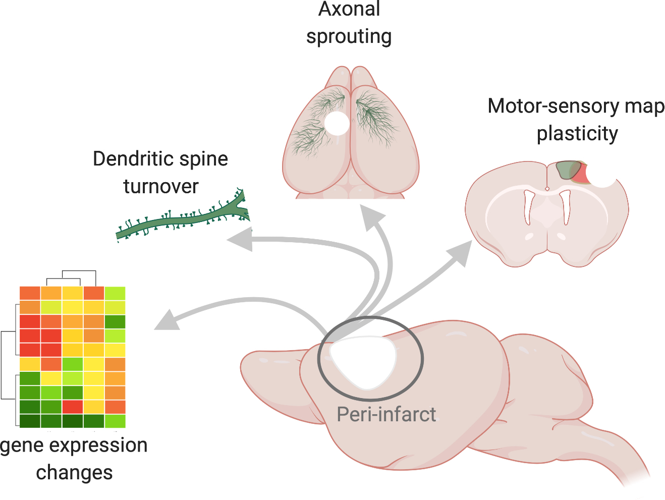
Sagittal view of a mouse brain (centre) in which the site of a stroke is depicted by the white area and the peri-infarct region is demarcated by a circle. The arrows point to molecular and cellular hallmarks of the peri-infarct region, including changes in gene expression. Here, these changes are denoted as a heatmap schematic in which the magnitudes of the changes in gene expression are visualized as differently coloured modules. New molecular programs support structural connectivity such as strengthening of connections through the growth and turnover of new spines, which are post-synaptic protrusions on dendrites, and the growth of new axons, depicted as blue fibres that sprout from the peri-infarct cortex and travel to intact motor, somatosensory and contralateral cortices. Changes in structural connectivity may underlie re-organization of functional motor (blue) and somatosensory (green) limb representations that re-appear at new locations several weeks after a stroke.