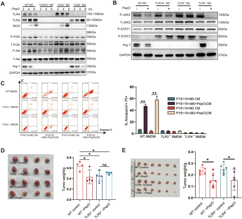
Fig. 5.
PepO reprogramed M2 macrophages via TLR2 and TLR4 recognition. A WT and TLR2 and/or TLR4 M2 BMDMs were stimulated with PepO(5 μg/ml), and samples were collected 2, 4, 6 h after stimulation. The phosphorylation of PI3K-AKT pathway associated proteins and M1 and M2 markers were evaluated by western blot. B WT and TLR2 and/or TLR4 M2 BMDMs were stimulated with PepO(5 μg/ml) for 24 h, The phosphorylation of JAK2-STAT3 pathway and the expression of Arg-1 was evaluated by western blot. C PY8119 cells were co-cultured with indicated CM for 48 h, and the percentage of apoptotic PY8119 cells labeled with PI and Annexin V were detected by FACS (n = 3). D, E TNBC was established in TLR2-/- (D, n = 4) and TLR4-/- (E, n = 6). Tumors were collected as previous schedule and the weight of tumor was measured. Two-way ANOVA with Tukey’s multiple comparisons test was used in (C). One-way ANOVA with Tukey’s multiple comparisons test was used in (D, E). Bar graphs represent mean ± SEM, *P < 0.05, **P < 0.01

