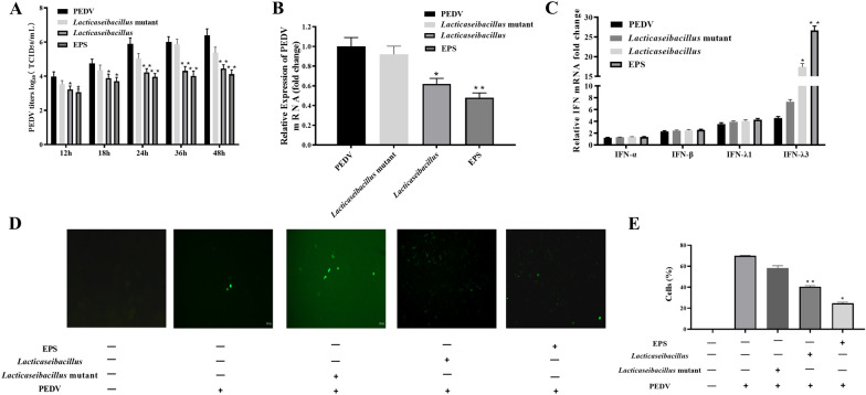
Fig. 4.
The antiviral effect of EPS, lacticaseibacillus and lacticaseibacillus mutant was analyzed by plaque assay with TCID50 in the IPEC-J2 in A. The TaqMan real-time RT-PCR was used to detect virus mRNA copies numbers in the PEDV replication in B. A standard curve was generated by plotting the threshold values against the serially diluted plasmid DNA encoding the PEDV M gene fragment. The C indicated that the type I and type III interferons, such as IFN-α, IFN-β, IFN-λ1 and IFN-λ3, expressed with the mRNA from the IPEC-J2 in the C. The indirect immunofluorescence results showed the PEDV distribution patterns detected in the IPEC with the rabbit polyclonal antibodies, which were used to analysis the EPS lacticaseibacillus and lacticaseibacillus mutant function against PEDV infection in the IPEC-J2 (D–E). The upper pictures are from the fluorescent (FITC) channel, and the bottom pictures are information added in the cell culture media in each groups. The results are represented as the mean ± SEM of three independent experiments. Data with * denote p < 0.05, ** denote p < 0.01

